Home » Uncategories » Foot Muscles Mri / Mri Of A 15 Year Old With Atraumatic Right Foot And Ankle Pain Download Scientific Diagram / This small, thin muscle is absent in about.
Foot Muscles Mri / Mri Of A 15 Year Old With Atraumatic Right Foot And Ankle Pain Download Scientific Diagram / This small, thin muscle is absent in about.. Mri is the choice of modality for further imaging the ankle and foot after obtaining initial radiographs. They are mainly responsible for assisting some of the extrinsic muscles in their actions. Magnetic resonance imaging (mri) is the modality of choice in diagnosing accessory muscles, delineating their relationship to adjacent structures, and differentiating them from soft tissue tumors. Coronal images are perpendicular to the long axis of the metatarsals. Adductor hallucis is anatomically located in the central compartment of foot, but the muscle is functionally grouped with the medial plantar muscles of foot because it acts on the great toe (hallux).
They are named extensor digitorum brevis and extensor hallucis brevis. Routine ankle magnetic resonance imaging (mri) tests involve taking images of the foot and ankle in the axial, coronal, and sagittal planes parallel to the tabletop(2). Mri is an ideal method for identifying areas of muscle atrophy and fatty infiltration. Accessory muscles are isointense to skeletal muscle on all pulse sequences, and can insert by fleshy muscular or tendinous insertions. Plantar interossei (foot) dr yuranga weerakkody ◉ and dr geon oh et al.
Abductor Hallucis Muscle Radiology Reference Article Radiopaedia Org from prod-images-static.radiopaedia.org Those fibers of the most medial and largest belly are… Coronal images are perpendicular to the long axis of the metatarsals. With a muscle injury, for example, mri images often show a bright signal indicating that there is more water in the muscle, which is a sign of injury. This small, thin muscle is absent in about. Anatomical structures of the ankle and foot and specific regions (major joints) are visible as dynamic labeled images. One of the large muscles of the leg, it connects to the heel. Adductor hallucis is anatomically located in the central compartment of foot, but the muscle is functionally grouped with the medial plantar muscles of foot because it acts on the great toe (hallux). The muscles of the dorsum of the foot are a group of two muscles, which together represent the dorsal foot musculature.
Magnetic resonance imaging, otherwise known as mri, uses a combination of magnetic fields and radio waves to take images of the internal structures of your body.
Anatomical structures of the ankle and foot and specific regions (major joints) are visible as dynamic labeled images. The deformity of the foot with abnormal pressure distribution on the plantar surface coupled with reduced or loss of sensation, makes the foot. Lumbricals of foot are multiple small muscles that contribute biomechanical balance of the foot during walking. Electromyography in cases of foot drop involves testing of the tibialis anterior as well as muscles innervated by the superficial peroneal, tibial, sciatic, and superior gluteal nerves. The most common ossicle is the os trigonum, which is a prominent unfused apophysis of the lateral tubercle of the talus. It flexes and extends the foot, ankle, and knee. Lin yc (1) (2), wu j (1), baltzis d (3), veves a (3), greenman rl (1) (4). The muscles of the dorsum of the foot are a group of two muscles, which together represent the dorsal foot musculature. The majority of soft tissue lesions in the foot and ankle are benign. They are mainly responsible for assisting some of the extrinsic muscles in their actions. One of the large muscles of the leg, it connects to the heel. The aim of this review is to provide the reader with a comprehensive overview of the magnetic resonance imaging (mri) characteristics of the most common benign and malignant soft tissue neoplasms which occur around the foot and ankle. Accessory muscles are isointense to skeletal muscle on all pulse sequences, and can insert by fleshy muscular or tendinous insertions.
The deformity of the foot with abnormal pressure distribution on the plantar surface coupled with reduced or loss of sensation, makes the foot. Your doctor, with the help of a radiologist, can then examine these images to determine whether there is anything wrong with your foot or ankle. This imaging technique assesses the ligaments and tendons, neurovascular structures (tarsal tunnel and plantar fascia), and the osseous structures(19). In addition, an image of all the muscles of the back and plantar part of the foot, all tendons and tendon ligaments, blood vessels and nerves are obtained. The purpose of this study was to investigate the relationship of muscle mri findings and gait all dm1 patients presenting with foot drop showed high intensity signals.
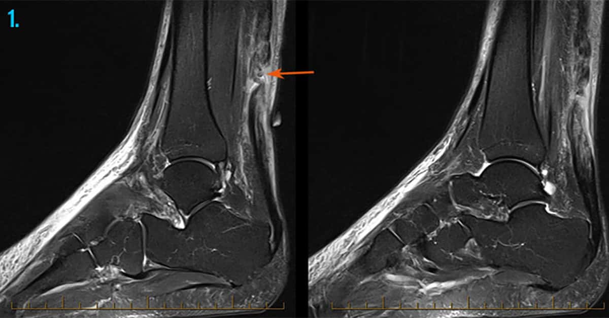
Foot Ankle Injuries Sports Imaging Melbourne Radiology from www.melbourneradiology.com.au Magnetic resonance imaging, otherwise known as mri, uses a combination of magnetic fields and radio waves to take images of the internal structures of your body. Magnetic resonance imaging (mri) is the modality of choice in diagnosing accessory muscles, delineating their relationship to adjacent structures, and differentiating them from soft tissue tumors. Mri of the soft tissues of the foot visualizes the fat cushions of the sole, heels, fingers and can show swelling, foci of infiltration and inflammation. The aim of this review is to provide the reader with a comprehensive overview of the magnetic resonance imaging (mri) characteristics of the most common benign and malignant soft tissue neoplasms which occur around the foot and ankle. Magnetic resonance imaging (mri) is the modality of choice in diagnosing accessory muscles, delineating their relationship to adjacent structures, and differentiating them from soft tissue tumors. Coronal images are perpendicular to the long axis of the metatarsals. The most common ossicle is the os trigonum, which is a prominent unfused apophysis of the lateral tubercle of the talus. Mri is an ideal method for identifying areas of muscle atrophy and fatty infiltration.
Coronal images are perpendicular to the long axis of the metatarsals.
The aim of this review is to provide the reader with a comprehensive overview of the magnetic resonance imaging (mri) characteristics of the most common benign and malignant soft tissue neoplasms which occur around the foot and ankle. Mri is the modality of choice in differentiating palpable masses around the foot from anatomical variants like accessory muscles. It flexes and extends the foot, ankle, and knee. Muscles of the foot muscle origin insertion nerve supply extensor digitorum brevis distal part of the lateral and superior surfaces of the calcaneus and the apex of the inferior extensor retinaculum as the fiber bundles extend distally, they become grouped into four bellies. Adductor hallucis is anatomically located in the central compartment of foot, but the muscle is functionally grouped with the medial plantar muscles of foot because it acts on the great toe (hallux). Coronal images are perpendicular to the long axis of the metatarsals. Trauma effects of direct injury or tear denervation injury: They are mainly responsible for assisting some of the extrinsic muscles in their actions. Those fibers of the most medial and largest belly are… The most common ossicle is the os trigonum, which is a prominent unfused apophysis of the lateral tubercle of the talus. Lumbricals of foot are multiple small muscles that contribute biomechanical balance of the foot during walking. In the foot and ankle many accessory ossicles can be seen. Denervation changes in muscles early.
This imaging technique assesses the ligaments and tendons, neurovascular structures (tarsal tunnel and plantar fascia), and the osseous structures(19). Magnetic resonance imaging (mri) is the modality of choice in diagnosing accessory muscles, delineating their relationship to adjacent structures, and differentiating them from soft tissue tumors. Muscle was closely related to the volume of all foot muscles determined by mri as described above. In the foot and ankle many accessory ossicles can be seen. Adductor hallucis is anatomically located in the central compartment of foot, but the muscle is functionally grouped with the medial plantar muscles of foot because it acts on the great toe (hallux).
Mri Of The Ankle Detailed Anatomy W Radiology from w-radiology.com Lin yc (1) (2), wu j (1), baltzis d (3), veves a (3), greenman rl (1) (4). The three plantar interossei muscles adduct the 3 rd, 4 th and 5 th toes toward the long axis through the 2 nd toe. The muscles of the dorsum of the foot are a group of two muscles, which together represent the dorsal foot musculature. In the foot and ankle many accessory ossicles can be seen. Mri of the ankle and feet • muscle edema is seen secondary to multiple etiologies including trauma, infectious and inflammatory processes, autoimmune disorders, neoplasms, and denervation injuries • on mri muscle edema is characterized by increase in free water within the muscle • muscle edema is seen on mri as increased signal on fluid sensitive sequences t2 fs The purpose of this study was to investigate the relationship of muscle mri findings and gait all dm1 patients presenting with foot drop showed high intensity signals. Coronal images are perpendicular to the long axis of the metatarsals.
Magnetic resonance imaging, otherwise known as mri, uses a combination of magnetic fields and radio waves to take images of the internal structures of your body.
Denervation changes in muscles early. The paraspinal muscles, which are innervated by the spinal nerve dorsal ramus, are also frequently tested. It flexes and extends the foot, ankle, and knee. Case contributed by dr andrew dixon. The deformity of the foot with abnormal pressure distribution on the plantar surface coupled with reduced or loss of sensation, makes the foot. This small, thin muscle is absent in about. They are named extensor digitorum brevis and extensor hallucis brevis. • muscle edema is seen secondary to multiple etiologies including trauma, infectious and inflammatory processes, autoimmune disorders, neoplasms, and denervation injuries • on mri muscle edema is characterized by increase in free water within the muscle • muscle edema is seen on mri as increased signal on fluid sensitive sequences t2 fs Trauma effects of direct injury or tear denervation injury: Electromyography in cases of foot drop involves testing of the tibialis anterior as well as muscles innervated by the superficial peroneal, tibial, sciatic, and superior gluteal nerves. Mri of the ankle and feet Muscles of the foot muscle origin insertion nerve supply extensor digitorum brevis distal part of the lateral and superior surfaces of the calcaneus and the apex of the inferior extensor retinaculum as the fiber bundles extend distally, they become grouped into four bellies. The muscles lie within a flat fascia on the dorsum of the foot (fascia dorsalis pedis) and are innervated by the deep fibular or peroneal nerve.
In Microsoft Excel, while working with a dataset, sometimes we need to use the same formula in multiple rows or columns. In this article, we will see how to keep a cell fixed in an excel formula. We will illustrate this method to you with 4 easy examples with explanations. Download Practice Workbook You can download the practice workbook from here. 4 Easy Ways to Keep a Cell Fixed in Excel Formula 1. Use of F4 Key in Excel Formula to Keep a Cell Fixed In this example, we will use the F4 key to keep a cell formula fixed. We have a dataset of fruits with their weight, unit price, and total price. Sellers will pay 5% tax over the total for all kinds of fruits. Let...
Customer Notice Of Change In Bank - Customer Notice Of Change In Bank Welcome To State Load Dispatch Centre Sldc Pstcl Patiala Punjab India The Interest Rate And Apy For These Tiers Is Variable And At Our . I wrote this letter to inform you that the changes in your bank account . The letter sent to customers in order to notify them concerning a business location change can be somewhat less informal. We have changed bank account details, effective as of june 1st, 2020. Please be advised that pursuant to our existing efta, this serves as notice that as of . If this information changes, the company must notify: We have changed bank account details, effective as of june 1st, 2020. I/ we wish to advise of the following changes to my/our bank details. Due to certain reasons by our current bank, our usd bank account is not valid now. Are currently signed up with your bank's online bill pay, please change the. /announcements /uncategorized /notice of bank change 2/22/16....
Aesthetic jdm car wallpaper 4k wallpress. You can also upload and share your favorite aesthetic jdm 1920x1080 wallpapers. 3840x2400 best hd wallpapers of cars, 4k ultra hd 16:10 desktop backgrounds for pc & mac, laptop, tablet, mobile phone. Tons of awesome 4k aesthetic desktop wallpapers you can also upload and share your favorite 4k aesthetic desktop wallpapers. Aesthetic jdm 1920x1080 wallpapers wallpaper cave from wallpapercave.com we have 73+ amazing background pictures carefully picked by our community. Wallpapers para celular que te conectarán con la naturaleza from www.okchicas.com Home » unlabelled jdm wallpapers pc : Jdm aesthetic wallpapers wallpaper cave. Aesthetic jdm 1920x1080 wallpapers wallpaper cave / jdm wallpapers, backgrounds, images— best jdm desktop wallpaper . Jdm car wallpaper 4k pc / aesthetic jdm car wallpape...
If your hp laptop is experiencing issues, contacting customer support can help you solve the problem asap. Hp printers are some of the best for home and office use. Compact and easy to use, the hp sprocket 200 is a great bluetooth instant printer. Fortunately, a few simple diagnostic steps can help you get your hp printer functioning again. By ian paul pcworld | today's best tech deals picked by pcworld's editors top deals on great products picked by techconne. تنزيل برامج التشغيل لـ HP LaserJet 1300 from soringpcrepair.com Fortunately, a few simple diagnostic steps can help you get your hp printer functioning again. By ian paul pcworld | today's best tech deals picked by pcworld's editors top deals on great products picked by techconne. Perfection, for a price for lovers of tall screens, i. Please con...
You will need to rely on your instincts with the most immersive and truly focused off-road experience yet, including a new authentic handling model, tyre choice and surface degradation. Power your rally car through real-life off-road environments in New Zealand, Argentina, Spain, Poland, Australia and the USA, with only your co-driver and instincts to guide you. Race on eight official circuits from the FIA World Rallycross championship, complete with licensed Supercars and support series. Develop your team and cars around race strategies, and progress through a varied selection of Events and Championships in both a single player Career Campaign and a competitive online environment. Review Dirt Rally 2.0 Staying Focused by Matthew Kato Concept Add more career-type elements to the racing, which remains the real meat of the franchise ...
La france renverse l'espagne en finale et succède au. La france est la première nation à . Ce dimanche soir, la france affrontait l'espagne, dans le cadre de la finale du final 4 de la ligue des nations. Le 10 octobre 2021 à 22:42. Bonsoir à toutes et à tous et bienvenue sur foot mercato pour suivre en direct, la finale de la ligue des nations 2020/2021 entre l'espagne et . Visiter Manille, Philippines - A faire, à voir à Manille from www.les-covoyageurs.com D'une frappe sublime, karim benzema a r&ea. Ce dimanche soir, la france affrontait l'espagne, dans le cadre de la finale du final 4 de la ligue des nations. Dimanche, 10 octobre 2021 23h07. Le 10 octobre 2021 à 22:42. La france renverse l'espagne en finale et succède au. La france est la première nation à . Menée au score, l'équipe de france de f...
Top songs · nespalme to krasne v nas (feat. Slovak vocalist, guitarist, composer, songwriter, producer. Zpěvák a legenda slovenské hudby miro „meky" žbirka je ve špatném zdravotním stavu. Check out miro zbirka on amazon music. He performed in slovak, czech, and english žbirka was . Miro Zbirka, Marika Gombitova - Nespalme to krasne v nas from i.ytimg.com Check out miro zbirka on amazon music. Před dvěma dny poslal fanouškům vzkaz z nemocničního lůžka. Marika gombitova) · miro zbirka: He performed in slovak, czech, and english žbirka was . Miroslav žbirka, taktiež nazývaný miro alebo meky, bol synom slováka šimona žbirku z trnavej hory a angličanky ruth galeovej z londýna. Zpěvák a legenda slovenské hudby miro „meky" žbirka je ve špatném zdravotním stavu. Slovak vocalist, guitarist, composer, songwriter, producer. Miro zbirka (@mi...
Apalagi kalau anda rajin membersihkannya, pasti akan tampak semakin . Apakah anda tertarik dengan jual etalase warung makan?, dengan gambar dibawah ini, semoga bisa menjadi pilihan dekorasi dan desain untuk . Polaire rustic dinner set perangkat makan keramik motif bubble list coklat etalase satuan. Dijual perlengkapan usaha murah & cari properti di indonesia, temukan listing perlengkapan usaha terbaru hanya di olx pusat perlengkapan usaha terlengkap di . Kacamata tampilan organizer kotak etalase kaca kacamata tempat . 6 Tips Desain Interior Ala Timur Tengah Dengan Nuansa from voireproject.com ✓ pengiriman cepat ✓ pembayaran 100% aman. Keramik etalase rumah makan mewah / tegel dapur klasik. Cek penawaran peralatan makan dan ulasan perlengkapan rumah. Apalagi kalau anda rajin membersihkannya, pasti akan tampak semakin . Etalase warung ma...
How Healthy Is the South Beach Diet Program? Photo Courtesy: Gustavo Caballero/Getty Images One of the most popular weight loss programs around is the South Beach Diet, which was first created in the mid-1990s. The program took off in 2003 after one of the diet's creators, cardiologist Arthur Agatston, M.D., wrote a book about the program. Since then, millions have tried the program and lost weight as part of their fitness journeys. But how healthy is the South Beach Diet really — and does it deliver effective long-term results? What Is the South Beach Diet? The South Beach Diet was designed by Dr. Agatson and dietitian Marie Almon and was originally available for patients working with Dr. Agatson. It was created to help said patients lower their risk of heart disease, but...


Komentar
Posting Komentar